Home
/ Horse Leg Bones Diagram - Forever Horses Anatomy Of The Equine Hindleg : Lameness problems from the fetlock down are usually caused by excessive.
Horse Leg Bones Diagram - Forever Horses Anatomy Of The Equine Hindleg : Lameness problems from the fetlock down are usually caused by excessive.
Horse Leg Bones Diagram - Forever Horses Anatomy Of The Equine Hindleg : Lameness problems from the fetlock down are usually caused by excessive.. Made up of the os coxae, the largest of the flat bones in a horse. This is logical, as a horse moves forward the bones of the lower leg shield the soft tissues. When the skeletal structure is properly proportioned the joints work smoothly. Learn vocabulary, terms, and more with flashcards, games, and other study tools. The suspensory apparatus, which carries much of the weight, prevents overextension of the joint and absorbs shock.
Learn vocabulary, terms, and more with flashcards, games, and other study tools. Finally, there is the large modern horse, equus, with only one toe, while all that is left of the other two are 'vestigial' splint bones. The shoulder joint is the articulation between the glenoid cavity of the scapula and the head of the humerus.in the horse, lateral and medial movements of this joint are impossible due to the shape of the humeral head; The limbs of the horse are structures made of many bones, joints, muscles, tendons and ligaments that support the weight of the horse's body. Horse leg bones diagram quizlet.
Horse Anatomy Leg Anatomy Drawing Diagram from shoestringstable.files.wordpress.com When the skeletal structure is properly proportioned the joints work smoothly. The shoulder joint is the articulation between the glenoid cavity of the scapula and the head of the humerus.in the horse, lateral and medial movements of this joint are impossible due to the shape of the humeral head; The arterial supply to the digit and fetlock of the thoracic limb comes mainly from the median palmar artery.the median palmar artery divides in the distal fourth of the metacarpus between thesuperficial and deep digital flexor tendons and the suspensory ligament, to become the medial and lateral digital arteries.part of the deep palmar arch anastamoses with the lateral digital. The extensor tendon attaches to the front of the coffin bone and straightens the leg; Their leg bones are proportioned differently from those of a human. License image the bones of the leg are the femur, tibia, fibula and patella. The ischium forms the point of the buttock. The pedal bone itself has an unusually high density of blood vessels within it.
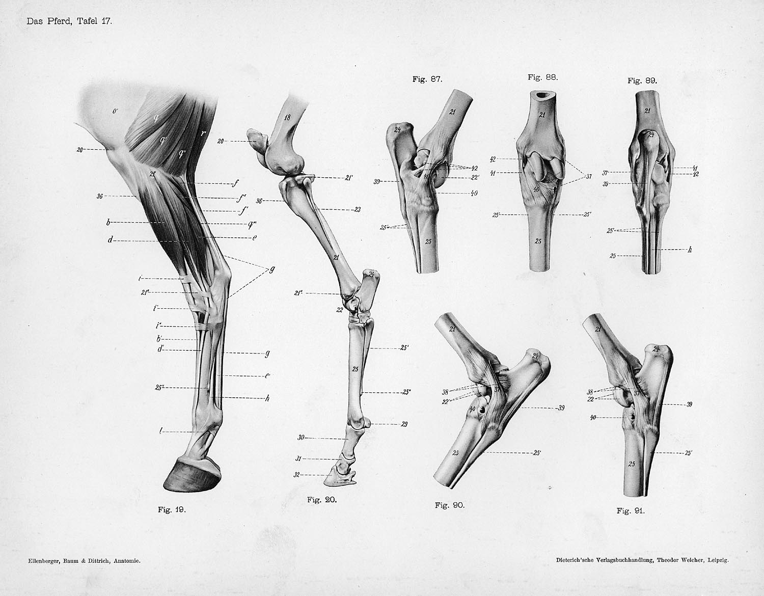
In this picture it shows the muscles that are closest to the surface of the skin, making them superficial.
We make sure to keep the original photos without changing. Horse leg bones diagram quizlet. Finally, there is the large modern horse, equus, with only one toe, while all that is left of the other two are 'vestigial' splint bones. The blood supply to and/or from the navicular bone is disrupted. The line leading from eohippus to the modern horse exhibits the following evolutionary trends: The ideal horse has legs which are straight, correctly set and symmetrical. This diagram shows the superficial layer of the tissue. The hoof is heavily supplied with blood through the two arteries which run down the back of the leg and into the foot. Individual horses may have structural defects, some of which lead to poor. In our website, we are persons who very treasure creativity from every one, with no exception. Their leg bones are proportioned differently from those of a human. Made up of the os coxae, the largest of the flat bones in a horse. Diagram of leg bones and joints via.
Horse body parts diagram, horse skeleton diagram and animal nervous system diagram are some main things we want to present to you based on the gallery title. The bones of the horse skeleton are held together with ligaments, tendons and muscles. The limbs play a major role in the movement of the horse, with the legs performing the functions of absorbing impact, bearing weight and providing thrust. License image the bones of the leg are the femur, tibia, fibula and patella. The bulk of soft tissue is behind the bones.
Limbs Of The Horse Wikipedia from upload.wikimedia.org The legs of a horse are made up of a system of muscles, tendons, ligaments, and connective tissue. Movement is therefore limited to flexion and extension. In the front of the leg, the only thing covering the bones is skin, the extensor tendons (which are very flat) and the suspensory ligaments and fascia. In this picture it shows the muscles that are closest to the surface of the skin, making them superficial. The extensor tendon attaches to the front of the coffin bone and straightens the leg; Hoof and lower leg structure. Talking about horse anatomy worksheets printable, we have collected some related images to complete your references. The blood supply to and/or from the navicular bone is disrupted.
The suspensory apparatus, which carries much of the weight, prevents overextension of the joint and absorbs shock.
The hoof is heavily supplied with blood through the two arteries which run down the back of the leg and into the foot. The pedal bone itself has an unusually high density of blood vessels within it. The suspensory apparatus, which carries much of the weight, prevents overextension of the joint and absorbs shock. Made up of the os coxae, the largest of the flat bones in a horse. See more ideas about horse. Learn vocabulary, terms, and more with flashcards, games, and other study tools. It is made up of the ilium, the ischium, and the pubis.at the junction of these three bones is a cavity called the acetabulum, which acts as the socket of the hip joint.the pelvic cavity is larger in diameter in the mare than in the stallion, providing more room for the foal during birth. If the angle at witch these bones are working is compromised, the joint becomes unevenly stressed and injury to the tendons and ligaments. They are joined to the spine through the sacroileac joints and allow transfer of propulsion to the hind legs. Few animals are as precocious as the horse. Principles of bone development in horses. The bones that make up the lower leg are the cannon bone, splint bones, long pastern, short pastern, pedal bone and navicular bone. Human anatomy muscles coloring pages printable via.
Horse body parts diagram, horse skeleton diagram and animal nervous system diagram are some main things we want to present to you based on the gallery title. Talking about horse anatomy worksheets printable, we have collected some related images to complete your references. Human anatomy muscles coloring pages printable via. In the front of the leg, the only thing covering the bones is skin, the extensor tendons (which are very flat) and the suspensory ligaments and fascia. This is logical, as a horse moves forward the bones of the lower leg shield the soft tissues.
The Horse S Skeleton Hind Limbs Youtube from i.ytimg.com The eohippus horses part b. Cow horse leg foot muscle and skeleton anatomy. The line leading from eohippus to the modern horse exhibits the following evolutionary trends: In this picture it shows the muscles that are closest to the surface of the skin, making them superficial. We make sure to keep the original photos without changing. This diagram shows the superficial layer of the tissue. This is supposed to demonstrate a change from browsing on bushes to grazing on grass. Similarly, the hock contains the bones equivalent to those in the human ankle and heel.
The shoulder joint is the articulation between the glenoid cavity of the scapula and the head of the humerus.in the horse, lateral and medial movements of this joint are impossible due to the shape of the humeral head;
The horses legs and hooves are also unique, interesting structures. Talking about horse anatomy worksheets printable, we have collected some related images to complete your references. Horse anatomy diagrams legs via. Within 20 minutes of birth a foal may stand, and within hours can be ready to run at speeds no human athlete will ever achieve. Cow horse leg foot muscle and skeleton anatomy. Horse body parts diagram, horse skeleton diagram and animal nervous system diagram are some main things we want to present to you based on the gallery title. The foot bones shown in this diagram are the talus, navicular, cuneiform, cuboid, metatarsals and calcaneus. Whereas, the deep digital flexor tendon runs down the back of the leg and wraps around the navicular bone, bending and flexing the leg. The pedal bone itself has an unusually high density of blood vessels within it. The limbs play a major role in the movement of the horse, with the legs performing the functions of absorbing impact, bearing weight and providing thrust. This is supposed to demonstrate a change from browsing on bushes to grazing on grass. The extensor tendon attaches to the front of the coffin bone and straightens the leg; The legs of a horse are made up of a system of muscles, tendons, ligaments, and connective tissue.
Hoof and lower leg structure leg bones diagram. Inflammation of navicular bone and/or bursa.